Posted on Leave a comment

Pine trees, conifers and evergreens


Quite frequently, someone I am enjoying time without outside will say something like “look over there by that pine tree.” Unfortunately, being the dork that I am, I am likely to look around and say “what pine tree?” Some of my friends have gotten clever enough to say “by that conifer”, or “by that evergreen”, thus releasing themselves from any obligation to get a tree identification perfect, and releasing me from dorkhood.

With that in mind, I decided to put together a little blurb on how to tell the different conifers apart. Fortunately, in the Valley, at least in the wild Valley, that isn’t too hard; there are really only 7 species in 5 genera to deal with… actual pines (2 species), true firs (1 species), Douglas fir (1 species), spruces (2 species) and juniper (1 species).

What they all have in common is needle or scale-like leaves, and (except for the juniper) reproductive structures that are obviously “pine cones”.

So here we go:

Pinus flexilis (limber pine) needle fascicle with 5 needles.

The actual pines, genus Pinus. Overall, there are 49 species of pines native to North America, but only three in the Valley (lodgepole pine, limber pine, and whitebark pine). They are easily told apart. As pines, all have needles borne in groups (fascicles), more or less joined at their bases.
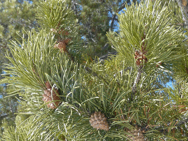
Lodgepole pine (Pinus contorta) branches

The most common numbers of needles are 2 (as in P. contorta) or 5 (as in P. flexilis and P. albicaulis). Up close, this is easy. From a distance, the fact that the needles are longer than other conifers makes them look more like bottle brushes.

Get used to the needles first, then stand back and look at the trees and where you are. Before long they will be so obvious to you, you will wonder what the problem ever was. (see also Pinus flexilisPinus contorta and Pinus albicaulis ).


Subalpine firs at Murphy Creek

The true firs, genus Abies. There are 7 species of Abies native to North America, but only 1 in the Valley. They are incredibly easy to spot from a distance because of their narrow, very pointed crown. If they happen to have cones, that too is a really obvious sign, in that unlike all the rest of the conifers, the cones stand up from the branches rather than hanging down.

Fir (Abies spp) needle cross section. The small hole in the lower middle is a resin duct. The bigger holes are intercellular air spaces.

The needles are also good clues; they are flattened so they don’t roll between your fingers. They are also connected straight to the twigs without petioles or pegs of any sort. And they aren’t pointy and sharp like spruce (see Abies lasiocarpa)
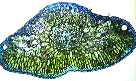

Douglas Fir needle cross section. Note the 2 resin ducts at either side (ends of the section). The blue stained tissue is heavily reinforced fibers for support and protection.

Douglas fir, genus Pseudotsuga. This not a true fir, but in a completely different genus. Like the fir, however, the needles are more or less flattened and won’t roll between your fingers.
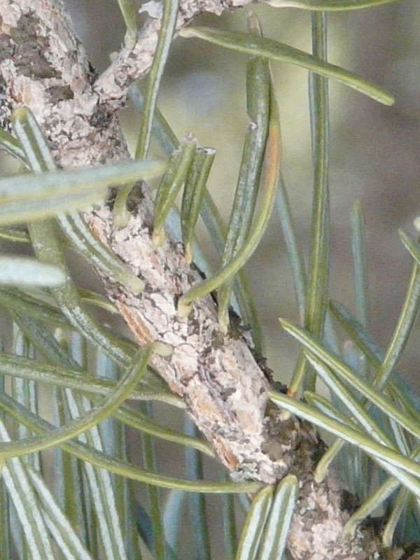
Douglas fir (Pseudotsuga menzeisiii) branch – note the way the needles are attached and the lines of stomates on the bottom sides

The needles are connected to the twigs by petioles (but no fascicles or pegs). The needle distribution on the twigs can be a bit confusing; they can either surround the twigs or be two-ranked (i.e. flat rows on opposite sides of the twigs). The twig buds are large, quite pointed and lustrous brown.
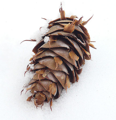
Douglas fir cone. Note the trident (mouse tail) bracts

What you can really rely on, however, is the cones, and you can often find some of these under the tree or on the ground nearby. The key is that they have trident (three pointed) bracts, like little mouse tails, sticking out from under the one scales. (see Pseudotsuga menzeisii).
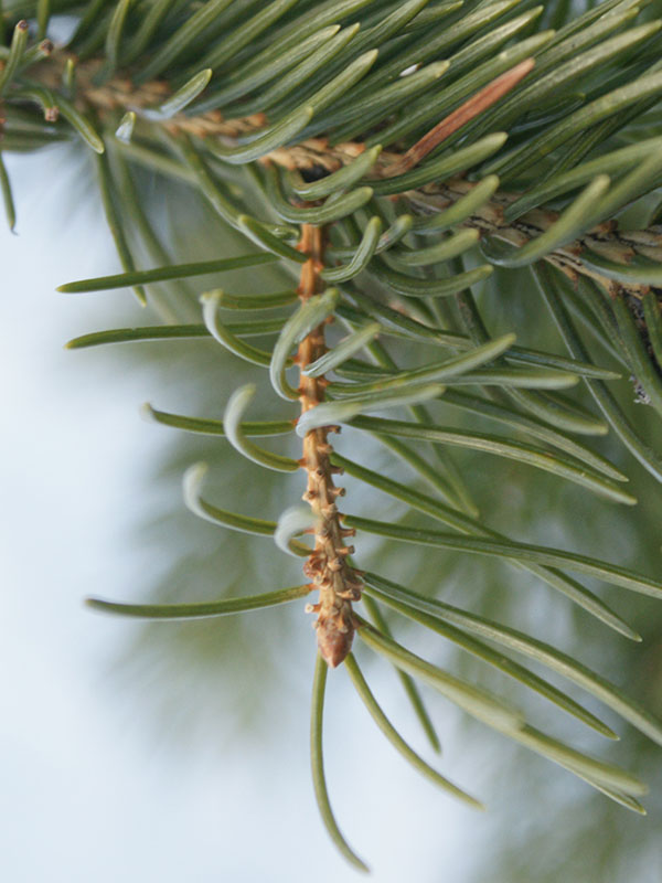

Blue spruce twig (Picea pungens) showing pegs (sterigmata).

Spruce, genus PiceaThere are two species of spruce in the Valley – blue spruce (Picea pungens) and Engelmann spruce (P. engelmannii) –  and they look a lot alike, even grading one to the other. Indeed, many authors note that not infrequently, it can be nearly impossible to distinguish between them. The Southwest Colorado Wildflower website, however, provides a good summary chart which I will use to give us all a better chance here.

First, however, the genus itself can be distinguished from the others Pinaceae by their needles and cones. Like the firs and Douglas fir, the needles occur singly, not in fascicles. In Picea, however, they are attached to the twigs by short, woody pegs (sterigmata). They are also uncomfortably pointy; blue spruce is downright awful.
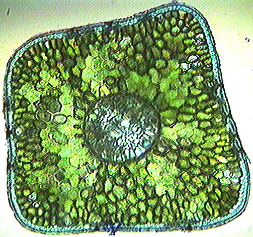
Spruce (Picea spp) needle. Note the fibers, and the square cross section.

In both species, individual needles are 4-sided and will roll between your thumb and fingers. The cones are at the top of the trees and are pendant (hanging downward). The scales are thin and completely unlike pine cones. And there are no mouse tails sticking out from under them.

To decide which of the two species you are looking at, try this:

  • Check the needles tips – while all spruce needles are somewhat spiky, the blue spruce needles are nastily sharp, Engelmann spruce less so
  • Needle color – totally unreliable and useless in distinguishing species
  • Blue spruce twigs are smooth while Engelmann spruce twigs are hairy. This may, however, only be discernible with a hand lens
  • Epicormic branching is rare in Engelmann spruce; the trunks are clean between branches. Epicormic branching is often extensive in blue spruce
  • Blue spruce trunks often have hard, vertically-ridged bark (and the epicormic branches);  Engelmann spruce trunks usually have flakey, scaley bark
  • Engelmann spruce is well adapted to higher and drier sites; blue spruce “likes” moister sites (stream sides) at lower elevations
Mixed spruce cones. The left most is Engelmann, the others are blue spruce.
  • Engelmann cones max out at about 2.5 inches long; blue Spruce cones start there. Engelmann cones fall off after seed maturity; blue Spruce cones hang on until the next spring (at least).
  • On older trees, Engelmann trunk bark is often cinnamon (orange) and scaled; blue spruce bark is gray and furrowed.

(see Picea engelmannii and Picea pungens)


Rocky mountain juniper (Juniperus scopularum) showing the berry-like cones and scale-like leaves.

Juniper, genus Juniperus. Finally, there is a single juniper species, J. scopularum, which is in the Cupressaceae rather than the Pinaceae and doesn’t look at all like a pine and doesn’t have “pine cones”. But it is still a conifer and still evergreen. Juniper leaves – at least past the seedling stage – are like overlapping scales rather than needles, and the cones look more like little blue berries. From a distance, junipers are conical with the branches often reaching to the ground… somewhat like the pseudo “pine” trees sold for model train sets. They tend to practice social distancing, i.e. the trees are usually not very close to one another. You’ll mostly see them on exposed, dry slopes along with mountain mahogany and limber pine. (see Juniperus scopularum.)


Posted on Leave a comment

Chara – should you care(a)?


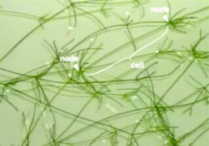
Fig. 1-Strands of Chara corallina. Each strand consists of large cells connected at nodes. The marked cell is approximately 7 cm long. (from PMC3592585, courtesy Bob Wyttenbach) Click to enlarge.

As a genus, Chara is cool. The plants look sort of like the flowering plants we celebrate generally on this site. But they are algae. Unlike the plants that are mostly emphasized on this site, Chara are also special because single “internodal” cells (stem lookalikes) can be huge. In Fig. 1 here, the single marked cell is about 7 cm long and 1 mm across! “Normal” plant cells are measured in micrometers.

So, what are these plants? Are they rare? Where did they come from?

Chara spp. are found world-wide, at least between 69˚N and 43˚S. But they are environmentally demanding, needing very clean water,  preferably with an above neutral pH. Interestingly, you may find either Chara or mosquito larvae in pond or stream water, but not both. Thus, the Teton River is a pretty good place for them. On the other hand, the main image in the gallery here for the species Chara contraria shows plants in a stock tank on a bike trail in Henderson Canyon.

OK, so I’ve identified the local species, but that doesn’t mean I know why. The identification of the genus to species is very difficult… depending on very small morphological features which are themselves quite variable. These days, charaphytologists (and yes, they actually call themselves that) are more likely to rely on DNA bar coding based on the matK gene–from chloroplasts and other plastids–to separate or group the species.

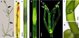
Regardless, all Chara species are multicellular algae with slender green or grey stems. Unlike familiar flowering plants, they don’t have apical meristems from which they grow. They just grow by cell division and specialization all over the plant. The grey color is cool because it results from lime (alkali) incrustations on the outer cell walls. These mask the green color of the chloroplasts. They are also rough feeling, hence the common name, stoneworts. Fig. 2 shows some details of this phenomenon, including that Chara actively raises the pH of the surrounding medium at specific locations on the cell surfaces. (It seems you can also keep Chara on agar, a sort of jello-like medium used widely in plant biology.)


Fig. 2. Thallus and internodal cells of Chara australis. a) The thallus (body) consists of internodal cells (arrows) separated by groups of smaller nodal cells (white arrow heads). Branchlets (a whorl of shorter internodal cells) extend from the nodes. b) pH banding pattern of an internodal cell visualized by phenol red (stain) in a solid medium. Pink color indicates alkaline pH. c), d) Calcified cell region in bright field (c) and in dark field microscopy (d). e) Tip of a thallus (dark field microscopy). Arrows show CaCO3 deposits “glueing” together the tips of branchlet internodal cells. f), g) CaCO3 deposits (arrows) between branchlet internodal cells from above (f) and from the side (g). Bars are 1 cm (a), 2 mm (b), 1 mm (c–e), 150 μm (f, g). Figure from Eremin et al. 2019 under creative commons license.

Getting back to the growth form of the alga, well before my time (i.e. in 1623), Characean algae were described as species of Equisetum owing to their superficial resemblance to horsetails (e.g. Equisetum arvense). Although they are not closely related at all, there is an evolutionary connection (see below). So at least superficially, the stoneworts resemble “land plants” in that they have stem-like and leaf-like structures. They also have something resembling roots… rhizoids.

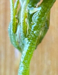
In nearly all Chara species, the internodal cell form the main axis. Often these are reinforced and stabilized by a layer of smaller cortical cells. Certainly, our local species, as shown (sort of) in Fig. 3, has these.

Chara contraria node
Fig. 3. Chara contraria – a node showing small cortical cells (sort of visible). Click to enlarge.

But this is not universal, and in particular, it is not true of Chara australis, Chara braunii, and Chara corallina (Fig. 1).

OK, so what? Is there anything worthy of attention besides this being a sort-of-unusual component of the local ecosystem?

Well, yes, there are a whole slew of reasons. For example, Chara has been an important organism for studying the evolution of what, for lack of a better word, we call “land plants”, or “embryophytes“, i.e. the plants most of interest to those looking at this website. Molecular studies of plant evolutionary relationships, “phylogeny“, have strongly suggested a close relationship between Characeans and those plants… perhaps not to the extent historically suggested that Characeans were the direct ancestor of “land plants”, but close enough. They share, for example,  a number of features–the fine structure and pigment composition of chloroplasts, and the composition and structure of their cell walls. (Cell walls are much more than cellulose, and even that isn’t found in all algae.)

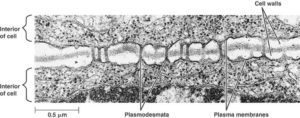
Plasmodesmata
Fig. 4. Plasmodesmatal connections between plant cells. Click to enlarge.

In addition, both groups have cell-to-cell connections through their cell walls, called “plasmodesmata” (Fig. 4). They share a unique mode of cell division, forming “cell plates” between the daughter cells. And they have similar growth controlling substances, a.k.a. hormones, not found in most algae.

For more on the origin of land plants, go here.

Well, and another reason to care…  Partly because of this close evolutionary relationship, these species have received a lot of attention by physiologists especially with respect to the transport of ions (nutrient) across membranes and various other membrane processes such as turgor, the pressure that keeps plants rigid (and is that is lost when plants wilt). Those membrane phenomena were what first got me started on liking Chara in the first place. I suppose I got sort of carried away and actually named my cat Chara.

The size of a Chara corallina (or C. australis) internodal cell, for example, and the fact that there are no “interfering” cortical cells around it, make It is easy to impale it with micro-electrodes. Those are very fine-tipped glass pipettes (tubes) that serve as electrical probes to study the voltage, currents, or even ion concentrations across cell membranes. Similar pipettes can be used to “perfuse” these cells, i.e. to pump fluids through them, completely replacing the cytosol and vacuolar contents. This is a handy manipulation if you want to see how transport varies with gradients across membranes. The National Library of Medicine currently lists 800 scientific articles about Chara corallina, many of which were based on just these techniques.

As a teaching tool, Chara corallina is also cool because it is easy to grow, and for demonstrations of phenomena ranging from action potentials (like nerves have, but slower) to growth, to the beautiful phenomenon of  cytoplasmic streaming (Fig. 5).


Fig. 5. Cytoplasmic streaming in a single Chara cell whose diameter (top to bottom) is approximately 1 mm. That’s huge. And the length is obviously much  much greater. The big blobs are oil droplets (probably) and occasionally you’ll see a paramecium swimming around because the sample was just “mounted” in stream water. From Jan-Willem van de Meent and Ray Goldstein via YouTube.


Finally, there’s the question of sex and reproduction. Sorry, but this could get confusing with all the specialized words. You can skip it if you like.

Like other algae, the main plant body of Chara is haploid. That means, unlike you and all the flowering plants we know, the cells have only one set of chromosomes. It’s called a gametophyte, because it can produce gametes (male and female cells). That’s fine if you want to reproduce only by cloning, but really, you want something like a “seed” for better dispersal and adaptation to different environments over time.

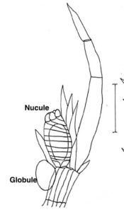
Fig. 6. A Chara contraria branchlet with a globule and nucule. From Ghazala et al. (2004) Pakistan Journal of Botany 36(4):733-743

And indeed, Characean reproduction does involve sex. The male gametes are produced in a sex organ called a globule (Fig. 6). The females are produced in nucules. In some species (including our local one), the two gametes organs are on the same plants; In that case, the males always develop first. In others species, they are on different plants.

Sexual reproduction results in a zygote that develops into an oospore. This isn’t a seed, per se but it serves the same purpose. The developing oospore, the only diploid stage of the life cycle, has an extremely hard, calcified cell wall which allows survival in wet or dry sediment for many years or even decades. Once good conditions have returned, the oospore cells undergo meiosis (cell division reestablishing the haploid state) as the plant grows into the easily-visible gametophyte.


A bonus feature – Back in the early 1980s, at a meetings in Toronto centered on plant membrane transport, a senior researcher from Adelaide, Australia – F. Andrew Smith– wrote new words to a popular song. To fully understand it, however, you need to be familiar with that song…  All around my hat … first recorded in 1976 by Steeleye Span.

In any case, here are the revised words:

All around the cell
There is a band of alkali
All around the cell
And it’s formed in the light
If any one should ask me
Just what it is that forms it
It’s formed by photosynthesis
And it goes away at night

Be well, all

Posted on Leave a comment

On pseudoflowers and parasites


This is the story of a plant and a parasitic fungus. The plant is a species of Boechera  – rockcress – in the Brassicaceae, i.e. the mustard family. The fungus is one that causes a disease called “rust”, Puccinia monoica. 

This is going to get weird and complicated, and may depend on you at least pretending to remember some of your high school biology, so to be easy on us all, I will start with the plant.

Boechera is a genus that includes a couple dozen species, most of which were formerly in the genus Arabis. Like many other small brassicas, rockcress’s main concern in choosing a habitat is to find a place where it doesn’t get drowned, and where it can get in and out before bigger things overtop it. For example, rocky habitats with shallow soil work well. Also like many brassicas, the rockcresses are by and large biennial. They put out a rosette of leaves in the first year, perhaps in late summer or early autumn, then “bolt” in the second year, putting on one to several erect stems with flowers at the tops in a tight and narrow inflorescence. In some cases, these can be as much as 3 feet tall, but generally they are much smaller.

In the Valley, I have seen this parasite only on what I believe to be B. pauciflora. For now, however, I rather like drawing on the left of one species from the 1912 edition of the Fieldbook of American Wildflowers by F. Schuyler Mathews. It is closely related…

Although B. pauciflora itself doesn’t have any special ecological role, plant molecular biologists are interested in boecheras partly because of their small genomes. B. stricta, for example, one of the species found in Idaho, has a genome of about 227 Mbases (that’s considered small) on 7 chromosomes (also small). It is one of two Boechera species whose genomes have been completely sequenced and it has been the subject of several hundred scientific articles… not bad for a small, ephemeral, nondescript, unimportant plant. There is an incredible paucity of information of any sort available on B. pauciflora.

Another very interesting feature of many species in this genus is that they reproduce asexually, producing seeds by apomixis… similar to the way dandelions make their seeds. Nevertheless, they do make pollen, and do attract insects to carry that pollen to other flowers. This is an important part of our story.

On the fungal side, Puccinia is one of about 168 genera of rust fungi; half of all the 7000 rust fungus species are in this genus, including our star, P. monoica. More broadly, all of these are in the larger group, the Basidiomycota, which also includes almost all the fungi (mushrooms) we intentionally eat. 

All the rust fungi are obligate parasites, and they have by far the most complex life cycles of any fungi, probably of any eukaryote. 

The Puccinia monoica life cycle, modified from N. P. Money (see source list). Chromosome configurations are shown as n, (n+n) etc.

The general life cycle of P. monoica is illustrated in the diagram to the right. It requires two unrelated plants, five stages, and five different kinds of spores, each with its distinct spore producing fruiting bodies. None of these is big enough to qualify as a “mushroom”. Of course, in biology, nothing is simple (not that a 5 part life cycle could be simple), and the rusts can be parasitized by other fungi and their spores eaten a number of species of insects (e.g. gall midges).

Being a basidiomycete, one of the kinds of spores Puccinia produces is a basidiospore. It only produces these on one of its hosts, and by convention, this is called the “primary host”. In this case it is a wild grass of some (unidentified) sort. This stage is a good one to consider the “beginning” of the life cycle, keeping in mind that cycles don’t actually have beginnings any more than bike wheels have ends. This works because the basidiospores are produced in the spring when rock cress growth is starting up again. The critical step is for the basidiospore to land on the Boechera and germinate. The funny little drawing at the top of the illustration depicts a spore (on the left) with a flattened “appresorium” growing out of it and an infection peg (on the right) penetrating the rock cress epidermis.

We’ll get back to how basidiospores are formed, but for now, one important bit of Puccinia genetics is that each has only a single copy of the fungal genome, i.e. it is haploid. We call this n. Our own personal cells, other than eggs and sperm, are diploid… they have two versions of each chromosome and are called 2n. Basidiospores also only have one nucleus so they are monokaryotic.

After the spore has germinated and the mycelium has penetrated into the leaf, it simply goes on growing and spreading, sucking up nutrients from the plant as it goes. That’s what parasites do.

Before finishing the rest of the fungal life cycle, let’s talk first about what else the spreading fungus does. To me as a plant biologist, these are especially cool bits of biology.

As they grow, besides sucking up all the nutrients, the mycelia massively alter gene expression in the plant. The result is that eventually the fungus neuters its host and stops it from forming flowers… and from ever producing seeds. In the language of population ecologists or evolutionary biologists, this makes the fitness of these individuals zero.  Instead, the fungus forces our little plant to make what are known as “pseudoflowers.” These are the yellow parts that fool you, at first, into thinking they have petals. Pseudoflowers are basically altered leaves that look and smell like real flowers. In fact, they look like real flowers in both the visible and ultraviolet wavelengths that insect pollinators see. Pseudoflowers also produce insect-attracting nectar-like substances.

Puccinia monoica (the yellow bumps are the fungal spermatogonia) on Boechera pauciflora

Also on each pseudoflower are hundreds of small cups (spermatogonia) that produce spermatia, a second kind of spore. Visiting insects get covered with these and carry them to other infected plants, to other spermatogonia. The bumps on the tops of the pseudoflower “petals” in the photos are the spermatogonia. They are also on the stems, but not on the bottoms of the “petals”.

Individual spermatia are neither “male” nor “female” but of compatible or incompatible “mating types”. At this point, they are also still monokaryotic and haploid. If, after dispersal by the insects, two compatible spermatia contact each other, they fuse, but the nuclei stay separate. This odd behavior means that the resulting mycelium is dikaryotic – each cell contains two dissimilar, haploid nuclei (called n+n, not 2n). Stick with me; this is cool.

Thus infected, next, the pseudoflowers give up the whole charade of producing pigments and the dikaryotic mycelia start into producing the next set of fruiting bodies and their spores. The structures are called “aecia” and the spores, aeciaspores. Often, to avoid crowding or competition, the aecia form on the opposite side of the “petals” and leaves from the spermatogonia. As I write this in mid-April in Teton Valley, aecia seem not yet to have been formed.

Aeciaspores are also dikaryotic (n+n) but everything about their gene expression for structure and function is different from the spermatia; they are configured specifically to carry the infection away from the rock cress to the primary host. In the Valley, they have to wait for this until the grasses start growing again which is some time after the rock cress. Aeciaspores are produced in huge numbers. As they are dispersed by the wind, this at least increases their chance of some finding a suitable host rather than blowing away and rotting in oblivion somewhere.

At this point, I won’t go into details about the grass host; most sources don’t actually suggest what it is other than “some species of grass”. But it is important to finish off this life cycle problem. As shown in the illustration above, the infection process for the grass again involves spore germination, appresorial growth, an infection peg and the gradual takeover of the leaves. It does not, however, involve more pseudoflowers. Instead, on the primary host, as it grows and steals nutrients, the fungus forms yet another spore-producing body, the uredinia which produces urediniospores. These are still dikarytotic (n+n). They are also capable of infecting other grass plants and do so as long as there are plants to infect. It is these structures that give the leaves the rusty color responsible for the name of the fungus and the disease it causes.

But, alas, all good things do come to an end, even for parasitic fungi. Autumn comes, the weather turns cold, the grasses prepare to die back to their roots and rhizomes, and the fungus knows when to call it quits. At that point, the uredinia legally change their names to telia, and start producing teliospores. Teliospores are different in that they can not infect any host, but have thick walls and are quite good at surviving the winter on the dead grass leaves. The dikaryotic cells give up their own charade and the nuclei fuse. We finally have something that is diploid (2n) and monokaryotic. And when spring comes, each of these germinates, producing a basidium. There, meiosis happens, four genetically different basidiospores form (assuring diversity and adaptability of the fungus), and the whole cycle continues.

The Puccinia/Boechera symbiosis is, IMHO, one of the more fascinating in local plants whose manifestation is visible. But I’m not sure if I would put it among the most fascinating of all parasitic arrangements. If, in some macabre way, you like this stuff, consider reading Parasite Rex by Carl Zimmer.


Sources and acknowledgements: 

First an acknowledgement: Special thanks to the iNaturalist community, in particular A. J. Wright and Bonnie Semmling who provided the identification of this “flower” as a fungus. A. J. Wright also identified the plant host.

Thomas Roehl, 2016, Order Pucciniales, the Rust Fungi, Fungus Fact Friday #130 … a fascinating set of fungal-centered articles, put out weekly online.

Cano, Liliana M et al. “Major transcriptome reprogramming underlies floral mimicry induced by the rust fungus Puccinia monoica in Boechera stricta.” PloS one vol. 8,9 e75293. 17 Sep. 2013, doi:10.1371/journal.pone.0075293  … a full blown scientific journal article, but bits of the abstract are generally understandable.

Nicholas P. Money, 2016, Fungal Diversity, in The Fungi (Third Edition).  The life cycle diagram was modified from here.

Carl Zimmer, 2000, Parasite Rex: Inside the Bizarre World of Nature’s Most Dangerous Creatures.

Posted on Leave a comment

How to identify the wild buckwheats (or not)

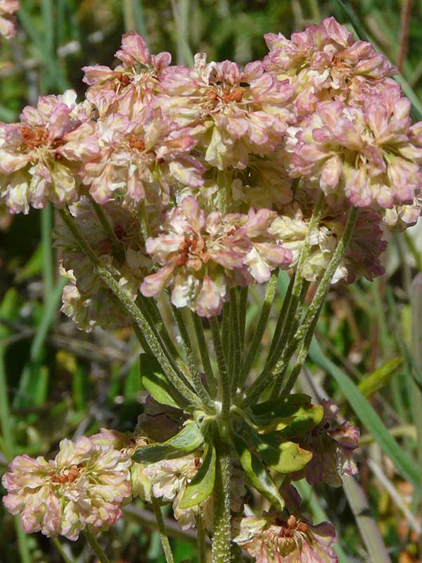
Wild buckwheat, seen from above

It is no particular trick to identify a plant as being a wild buckwheat, even in the winter. Mostly, you’ll see it from above, so looking down, you see cute little buttons that look sort of like dried flowers.  The buttons come in clusters of their own. You find them on dry hillsides or the tops of hills and mountains. And you’ll probably be happy about that.

Wild buckwheat leaves in winter

In the pre-snow winter, the leaves are strikingly red and green, quite beautiful on an otherwise taupe hillside.

Without too much effort, if you lie down on the ground or pick a stem and hold it up, you’ll see that all these flower clusters are actually at the tops of short pedicels as part of an umbel, and where they all join, there is probably a whorl of green, leaf-like bracts.

… wild buckwheat – note the whorl of bracts just below the inflorescence

So all this is easy and can be done in a minute. But that’s where it ends.

Start with color. The blossoms may be white, cream or red (all -ish). But… individual clusters may turn from white to pink and red as they age. So, looking at a flower cluster doesn’t really tell you what color to match it with.

And those bracts (the involucre). If you hold the blossoms upside down, you might (or might not) find another set below each individual cluster.

So what’s the problem? Earle and Lundin illustrate only 9 species, and they look different enough. Dee Stickler shows 3 species, all very different, in his Alpine book and 2 in his Prairie book.

The problem is, first, that Eriogonum is by far the largest genus in the family Polygonaceae. It includes some 230 species, mostly of western North American plants. The Idaho Department of Fish and Game lists 50 species present in Idaho. And second, let’s consider just one of those, sulphurflower wild buckwheat, Eriogonum umbellatum. As Earle and Lundin note in their book, this species is highly variable, with 40 or so recognized varieties, several of them, at least, common in Idaho. Or third, the fact that the species hybridize with each other, and otherwise have huge variations in form and color. Or fourth, that the genus is still undergoing speciation, i.e. many of the species themselves are not “stable”.

As the Southwest Colorado Erigonium page says: “Various Eriogonum species may be tall and lanky; matted; sprawling; bushy; herbaceous or woody; pink, white, or yellow flowering; annual, biennial, perennial, or monocarpic; and often in hot and dry environments.” That pretty much covers it. Well, and that they may have simple or compound umbels, absent or invisible involucres below the flower clusters, and … whatever.

Finally, even the descriptions of individual species published by different groups are so different it is hard to believe they are looking at the same plants… and their accompanying photos verify that. Earle and Lundin, for example, describe Erigonium pyrolifolium as having “smooth, bright green basal leaves and white flowerheads.”  The Wikipedia entry says it has woolly leaves and small hairy flowers ranging from greenish-white or white to pink. At Crater Lake, it is small and shrubby. pnflowers.com says the leaves can be hairy or smooth. The Jepson herbarium says they are smooth. The Montana field guide says they are “white- to brown-tomentose [hairy] below, nearly glabrous [smooth] above”. You get the message.

So the answer in this case is not 42, but that identification is really hard because there is just so much variability. Without a hand lens and a botanical key, and probably years of experience, real identification to species may be impossible. And it is certainly not going to happen (for most of us) in a minute. I’m happy to call it buckwheat.

ps: In case you wondered, wild buckwheats and culinary buckwheats are different. The culinary version is Fagopyrum esculentum, but is at least in the same family as the eriogonums.